
![[PS1000] FluoroStain™ Protein Fluorescent Staining Dye (Red, 1,000X), 1 ml](/web/image/product.template/340/image?unique=1c54916)
![[PS1000] FluoroStain™ Protein Fluorescent Staining Dye (Red, 1,000X), 1 ml](/website/image/product.template/340/image/90x90)


Description
The FluoroStain™ Protein Fluorescent Staining Dye (PS1000/PS1001) is a less toxic and more environment-friendly reagent and designed to substitute the Coomassie Blue protein staining method and Silver Stain, offering great sensitivity and ease of operation. Unlike the Coomassie Blue stain, the FluoroStain™ binds to proteins with high specificity and exhibits low affinity to polyacrylamide gel, making destaining an option rather than a requirement. With further reduction of background signals via destaining process, the FluoroStain™ Protein Fluorescent Staining Dye is capable of achieving detection levels parallel to silver staining. The FluoroStain™ is also compatible with mass spectrometry analysis, i.e. LC-MS/MS, MALDI-TOF etc., facilitating the use for proteomics analysis.
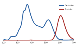
The FluoroStain™ Protein Fluorescent Staining Dye can be excited by UV and blue light sources, with excitation peaks around 369 and 517nm and emission at 605 nm. The FluoroStain™ is compatible with both ultra violet illumination systems and less harmful blue light illumination system, such as B-BOX™ Blue light epi-illuminator is recommended.
Spectral Characteristics
When it is bound with bovine serum albumin (BSA), the fluorescent emission of FluoroStain Protein Fluorescent Staining Dye can be excited by UV and blue light sources, with excitation peaks around 369 and 517 nm and emission at 605 nm. In absence of BSA, FluoroStain Protein Fluorescent Staining Dye shows ignorable fluorescence as compared with protein-bound form, therefore giving a clear background for photographic analysis.
These spectral characteristics made this fluorescent dye compatible with a wide variety of gel reading facilities, including UV/ blue light epi- and transilluminator, argon laser and mercury-arc lamp excitation gel scanners.
Storage
Protected from light
-20°C for 24 months
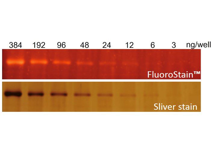
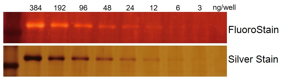
Sensitivity of FluoroStain™
Comparison of FluoroStain™ Protein Fluorescent Staining Dye (Red, 1000×) with Silver stain of a 2× serially diluted BSA sample.
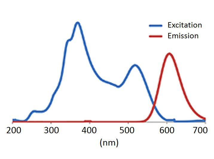
Spectrum of FluoroStain™
Excitation and emission spectrum of FluoroStain™ Protein Fluorescent Staining Dye (Red, 1000×).
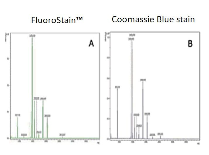
Compatibility to MASS analysis
Comparison of MALDI-TOF mass spectra of bovine serum albumin (BSA) stained with FluoroStain Protein Fluorescent Staining Dye (A) and with Coomassie Blue (B). BSA proteins are seperated on SDS-PAGE, stained with fluorescent dye or conventional Coomassie Blue, followed by trypsin digestion in gel, and then analyzed by MALDI-TOF.
Contents
| |||||||||
Can the SDS-PAGE gel stained with FluoroStain™ Protein Fluorescent Staining Dye (PS1000) be transferred to perform Western Blot? Will it affect the antibody binding when Western Blot is performed?
After using FluoroStain™ Protein Fluorescent Staining Dye (PS1000) to stain the gel, it is NOT recommended to perform Western Blot. The proteins in the SDS PAGE gel are fixed during staining with acidic solution, and thus preventing the transfer of proteins to NC or PVDF membrane.
When doing a follow up analysis using an SDS PAGE stained with FluoroStain™ Protein Fluorescent Staining Dye (PS1000), will there be any effect on mass analysis?
There will be no effect on mass analysis when following up with an SDS PAGE analysis using FluoroStain™ Protein Fluorescent Staining Dye (PS1000).
Is FluoroStain™ Protein Fluorescent Staining Dye (PS1000) able to stain a 2D gel?
The FluoroStain™ Protein Fluorescent Staining Dye (PS1000) can stain a 2D gel, which is quite sensitive. PS1000 is capable of achieving detection levels parallel to silver staining. The sample gel can be used for further MASS analysis.
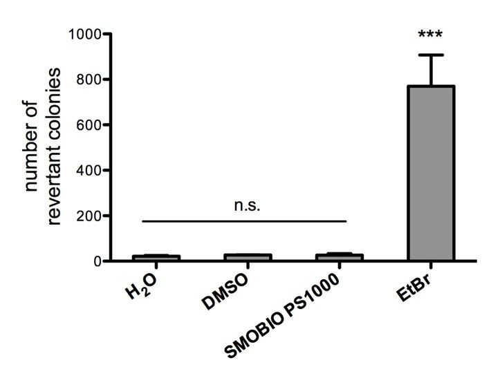
Are there relevant document that support safety (ex. non-mutagenicity) of FluoroStain™ Protein Fluorescent Staining Dye (PS1000)?
FluoroStain™ Protein Fluorescent Staining Dye (PS1000) is proofed for their safety (non-mutagenicity) using Ames test (Figures below). However, it must be noted that since solvent may penetrate the skin, it is recommended that users wear gloves when using the fluorescent dyes.
Working Reagent Preparation
1:1000 dilution in 40% ethanol and 2% H3PO4
Suggested volume of staining solution
|
Gel dimension |
Gel volume |
1× staining solution |
|
9 cm × 7 cm |
≈ 6.5 ml |
≈ 100 ml |
|
13 cm × 9 cm |
≈ 12 ml |
≈ 180 ml |
|
16 cm × 16 cm |
≈ 26 ml |
≈ 390 ml |
|
26 cm × 23 cm |
≈ 60 ml |
≈ 900 ml |
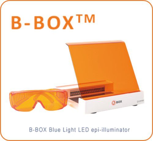
B-BOX™ Blue Light LED Epi-illuminator
470 nm long wavelength
Improved cloning efficiency
Compact, light-weight, and portable (less than 1 kg)
Adjustable and removable filter plate allows for gel cutting, visualization, and documentation
470 nm long wavelength
Improved cloning efficiency
Compact, light-weight, and portable (less than 1 kg)
Adjustable and removable filter plate allows for gel cutting, visualization, and documentation
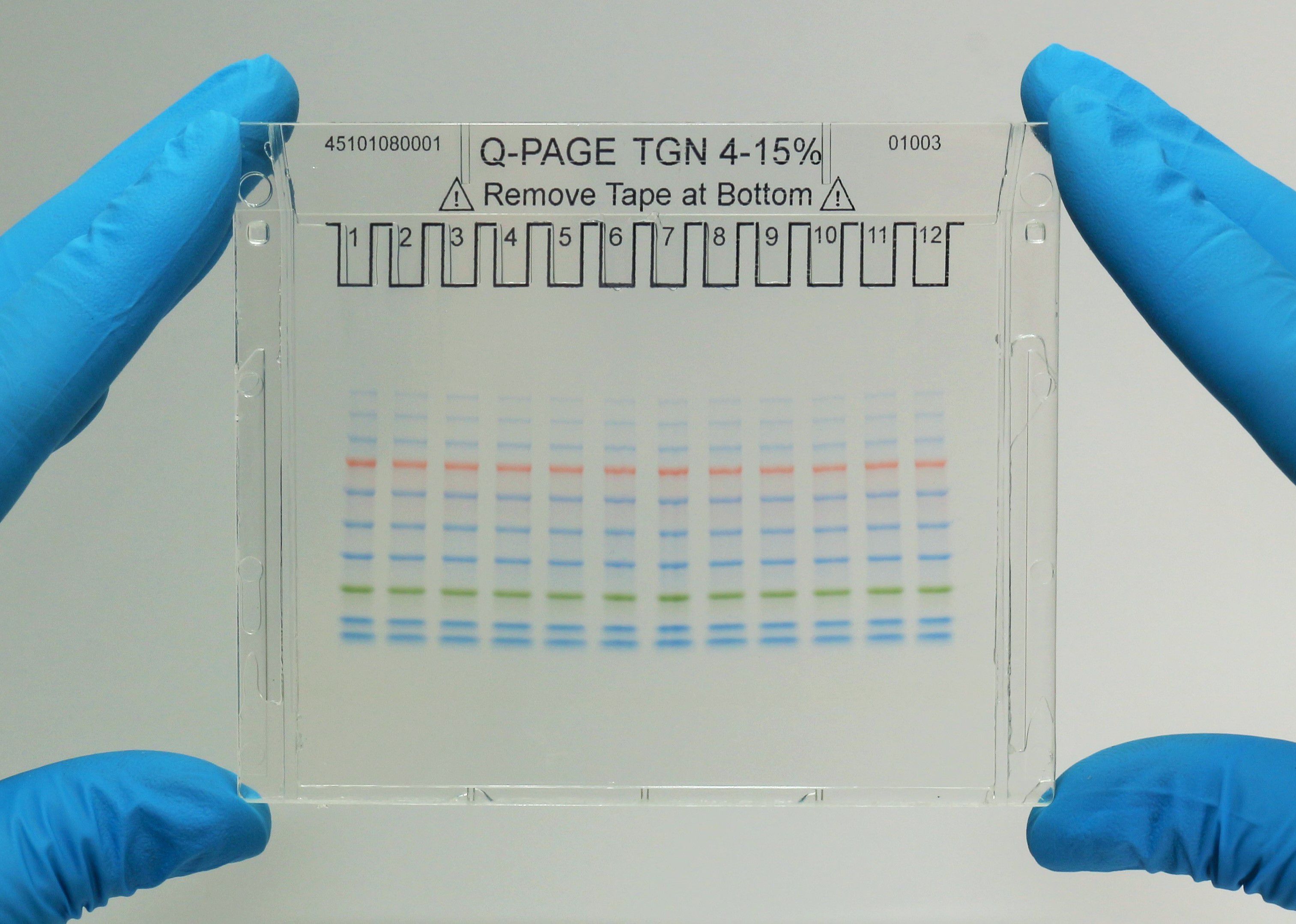
Q-PAGE™ Precast Gels
User-friendly gel cassette:
Numbered and framed wells for sample loading
Labeled warning sign and green tape as reminder
Enhanced gel performance:
Enhanced gel electrophoresis speed
Better band separation
Stable for shipping at ambient temperature
Easy compatibility:
Available as homogeneous and adjusted gradient gels for a wide range of protein separation.
Compatible with most popular protein electrophoresis systems
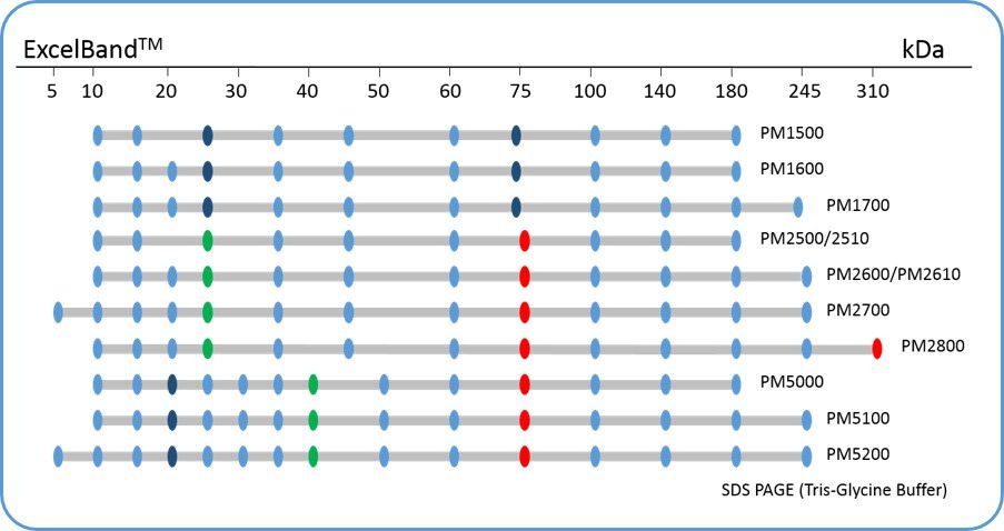
ExcelBand™ Protein Markers
Ready-to-use— premixed with a loading buffer for direct loading, no need to boil
Broad range— 310 kDa to 5 kDa
Pre-stained bands — for monitoring protein separation during electrophoresis and Western blotting transferring efficiency on membrane
Enhanced bands— for quick reference